The Nobel Prize in Physiology or Medicine was awarded for the study of the brain through light-gated ion channels and optogenetics. To honor that achievement, we are dedicating this blog to the past and future of optogenetics — how it works, how the tools have grown, and how it pairs with behavioral research today.
Researchers know a great deal about what different parts of the brain do. The harder question is which individual brain cells are responsible for a particular action or response. Optogenetics helps them investigate that connection by using light to change the activity of selected cells.
Traditional methods of brain research
For many years, researchers relied mainly on drugs and electrodes to study how brain cells affect behavior. Drugs can target specific receptors, but they take time to work and their effects can last longer than an experiment needs. Electrodes act quickly, but the electrical stimulation can also reach nearby cells that weren't meant to be involved.
What researchers needed was a way to control specific cells at a particular moment, more like flipping a light switch.
How light gives researchers more control
Scientists found a way to introduce light-sensitive proteins, originally discovered in algae, into mammalian neurons, or nerve cells. These proteins, called opsins, respond to certain wavelengths of light. They allow researchers to turn activity in selected neurons on or off, with much finer control over where and when that happens.
The technique offered a level of precision that earlier methods could not match. Therefore, Nature named it Method of the Year in 2010. This combination of light and genetics is called optogenetics.
"Optogenetics is a technology that allows targeted, fast control of precisely defined events in biological systems as complex as freely moving mammals."
Early experiments used tissue studied outside the animal. Researchers then moved to freely moving animals with implanted optical fibers that delivered light to the brain. The tools have continued to develop since those early studies. The discoveries that optogenetics has enabled have not gone unnoticed: Karl Deisseroth (an EthoVision user), Peter Hegemann, and Georg Nagel were awarded the Nobel Prize in Physiology or Medicine in 2026 for their work on optogenetics.

How the tools have developed
Why color matters, and how it has changed over the years
The first optogenetic tools used blue light. It scatters and is absorbed quickly in tissue, making it difficult to reach neurons more than a few hundred microns below the surface. That meant placing an optical fiber close to the cells being studied.
Newer, widely used opsins, including ChrimsonR and Jaws, respond to red light. These are known as red-shifted opsins. Red and far-red light, at wavelengths of roughly 620–700 nanometers, travels farther through tissue and bone and causes less light-related damage. Jaws, for example, can suppress neural activity up to 3 millimeters deep in the mouse brain through an intact skull.
Using more than one color also gives researchers more control. This approach is called multiplexing. By placing a red-sensitive protein that activates neurons and a blue-sensitive protein that suppresses them in the same brain circuit, researchers can switch between the two effects by changing the light's color. They can test both directions of control in the same animal, without changing the genetic setup.
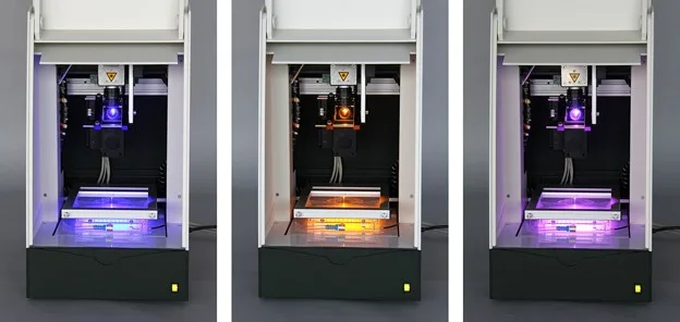
Optogenetic setup in the DanioVision system. Juvenile zebrafish can be exposed to different wavelengths of light.
CaMPARI: marking the brain cells that were active
Some light-based tools help researchers identify which neurons were active during a behavior, rather than change their activity. CaMPARI is one example. Developed at HHMI's Janelia Research Campus, it leaves a record that researchers can examine later, without keeping a microscope near the animal during the experiment.
Its full name is Calcium-Modulated Photoactivatable Ratiometric Integrator. That sounds technical, but the idea is straightforward: active cells are marked by a color change.
CaMPARI is a protein that cells are genetically instructed to make. It normally glows green. When a neuron is active, calcium levels inside it rise. If the cell is also exposed to violet or ultraviolet (UV) light at around 400 nanometers, CaMPARI permanently changes from green to red. This change is called photoconversion.
Researchers apply the light during the behavior or event they want to study. Only neurons that are active during that window turn red. The ratio of red to green reflects the calcium level, giving researchers a measure of activity rather than just an on-or-off label. Because the color change is permanent, they can examine the tissue afterward and map activity across the whole brain at the level of individual cells.
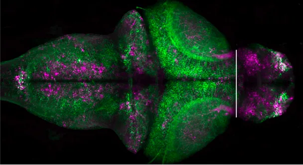
CaMPARI 2.0 displayed in a zebrafish larva. Image by HHMI Janelia.
This separates the behavior from the microscope work. Recent studies have used CaMPARI2 to map brain-wide activity in freely moving zebrafish larvae startled by a sound, and to mark active neurons in adult fruit flies during an unpleasant stimulus. Holding the animals still for imaging would have interfered with the behaviors being studied.
There is a catch: the light used to mark the cells can affect behavior, too. Zebrafish larvae can see UV light and may avoid it. Researchers therefore need to account for the possibility that the marking light itself becomes part of what the animal is responding to.
CaMPARI in rodents
The first tests of CaMPARI in living animals used rodents, and rodent studies remain an active area of research. In early mouse studies, researchers used it in the visual cortex, the part of the brain that processes visual information. They measured how strongly individual neurons responded to visual patterns at different angles. The amount of color change matched the strength of each response, showing that CaMPARI could capture differences in activity.
Since then, mouse studies have used CaMPARI to map working connections between brain regions and to study communication between neurons and astrocytes, another type of brain cell. Researchers have also recorded activity across large volumes of brain tissue while mice moved freely, without having their heads held in place or carrying a tether or miniature device.
In that approach, researchers shine the marking light over the mouse and its home environment while it performs behavioral and cognitive tasks. They examine the preserved tissue afterward with a two-photon microscope. Separating the experiment from the imaging made it possible to study a larger volume of the cortex than is practical with live imaging. The work revealed task-related differences in activity across areas involved in movement and in processing touch and body sensations.
CaMPARI can also be combined with optogenetic stimulation to map connections in freshly prepared slices of mouse brain tissue. Researchers use channelrhodopsin, a light-sensitive protein, to stimulate the ends of nerve fibers coming from one brain region. CaMPARI in a second region then marks the neurons that respond to those signals. In this setup, one light source both stimulates the first group of cells and marks the responding cells downstream — so researchers can identify those connections without recording electrical signals from the cells.
Innovations with and within optogenetics
In a simple behavioral experiment, entering a particular area can trigger a light pulse. EthoVision's Trial & Hardware Control can do this, as the place-preference example below explains. The same idea now extends to wireless implants and more detailed tracking of what an animal is doing.
This is often called closed-loop control: the system detects a behavior or change in brain activity and uses that information to decide when to stimulate. AI-guided tools can help identify the movements or postures that trigger the light.
- Stimulation triggered by movement or posture. Real-time tracking tools such as EthoVision, and pose-estimation tools such as DeepLabCut-Live, are routinely paired with stimulation hardware. Depending on the setup, a body or limb position, walking pattern, or posture can trigger the light.
- Experiments with multiple animals. Some systems track multiple animals and trigger stimulation during a particular social interaction. Researchers can reinforce or interrupt that interaction as it happens to study group behavior and social hierarchy.
- Fully wireless hardware. Soft, implantable probes with tiny LEDs remove the need for a tether. Some systems also include tracking, allowing stimulation and behavior measurement without a physical connection to the animal.
The underlying question is still the same: how does a particular change in brain activity affect behavior? These tools give researchers more precise control over the trigger and automate more of the experiment.
Optogenetics and EthoVision
EthoVision combines behavior tracking with programmed signals that control stimulation. Several long-established experiments show how this works in practice.
Real-time place preference: does the animal seek out or avoid stimulation?
In a real-time place preference test, an animal explores a rectangular arena with two distinct sides. Entering one side triggers optogenetic stimulation. Activating or suppressing the targeted neurons can make that experience rewarding or unpleasant.
Researchers then look at where the animal chooses to spend its time across sessions. Spending more time on the stimulated side suggests a rewarding effect; avoiding that side suggests an aversive, or unpleasant, effect.
EthoVision's Trial & Hardware Control can link the chosen zone to a trigger. A signal passes through the input/output (IO) box to switch on the light source. Researchers can also define several zones, each linked to a different stimulation frequency or light source.
Testing effects on anxiety-related behavior
Researchers can use a similar approach to test whether stimulating a group of neurons reduces or increases anxiety-related behavior. For example, an elevated plus maze test can use an off-on-off, or ABA, design: stimulation is off at the start, on for a set period in the middle, and off again at the end. If the animal spends significantly more time in the open arms during stimulation, the result suggests an anxiety-reducing effect. More time in the closed arms points in the opposite direction.
Free Trial
Want to run optogenetic experiments with precise, automated triggers?
EthoVision's Trial & Hardware Control links behavior tracking to light delivery, so stimulation happens exactly when your experiment calls for it. Request a free trial and see how it works with your setup.
Try it now!
arrow_forward
Movement and learning tasks
Researchers can also measure how stimulation changes movement in an open field, including speed, overall movement, and turning. These measures help connect the activity of particular neurons with what the animal does.
Triggers do not have to depend on location. Trial & Hardware Control can also respond to detected actions, such as a nose poke or lever press. This can automate operant learning tasks, in which an animal learns that an action leads to a rewarding or unpleasant outcome.
Where optogenetics goes from here
Optogenetics has become a standard tool for studying how brain circuits work, rather than the emerging technique it was in 2012. Its uses are still expanding.
Red-shifted opsins and experiments using more than one light color have made it routine to reach deeper tissue and control activity in both directions with less invasive methods. CaMPARI adds a way to identify which cells were active during a behavior. Wireless systems, closed-loop control, and AI-guided behavior analysis make it possible to automate experiments that once depended on manual triggering.
Research funding continues to support this work. The NIH BRAIN Initiative, the largest U.S. funder of neurotechnology tool development, received an additional $1.5 billion through 2026 under the 21st Century Cures Act. Its recurring funding calls include new tools for recording and changing neural activity, including optogenetics.
For researchers planning a place-preference test or an experiment with several zones and stimulation frequencies, EthoVision offers a way to link behavior tracking with light delivery. Trial & Hardware Control handles the programmed triggers, so stimulation happens when the experiment calls for it.
References
- Deisseroth, K. Optogenetics. Nat. Methods 2011, 8, 26–29.
- Hsueh, B.; Chen, R.; Jo, Y.; et al. Cardiogenic control of affective behavioural state. Nature 2023, 615, 292–299.
- Yang, Y.; Wu, M.; Vázquez-Guardado, A.; et al. Wireless multilateral devices for optogenetic studies of individual and social behaviors. Nat. Neurosci. 2021, 24, 1035–1045.
- Hong, S. Wireless optogenetic microsystems accelerate artificial intelligence–neuroscience coevolution through embedded closed-loop system. Micromachines 2025, 16, 557.
- Stamatakis, A.M.; Stuber, G.D. Activation of lateral habenula inputs to the ventral midbrain promotes behavioral avoidance. Nat. Neurosci. 2012, 15, 1105–1107.
- Peng, X.; Mao, Y.; Tai, Y.; et al. Characterization of anxiety-like behaviors and neural circuitry following chronic moderate noise exposure in mice. Environ. Health Perspect. 2023, 131, 107004.
- Stuber, G.D.; Sparta, D.R.; Stamatakis, A.M.; et al. Amygdala to nucleus accumbens excitatory transmission facilitates reward seeking. Nature 2011, 475, 377–380.