Fear Conditioning: How EthoVision XT Supports Your Behavioral Research
Last updated: Jul 24, 2026
Fear Conditioning: How EthoVision XT Supports Your Behavioral Research
Fear Conditioning: A Deeply-Rooted Behavior
Fear conditioning is a fundamental behavioral process seen across a variety of species, from rodents to humans. It is a classic form of Pavlovian learning where subjects associate a neutral stimulus (like a light or tone) with an aversive event (mild foot shock).
There are two widely used variants of fear conditioning:
- Cued fear conditioning, which links a stimulus with a shock;
- Contextual fear conditioning associates the environment with an unpleasant stimulus.
The most used readout for fear conditioning is freezing, which can be easily measured with the Ugo Basile Fear Conditioning System in combination with EthoVision XT [1-5].
Creating automated tests and reliable detection of freezing behavior are essential for reproducibility of your results. Noldus EthoVision XT combined with the Ugo Basile Fear Conditioning System provides a solution that is centered on just those two aspects.
How to Perform a Fear Conditioning Test
To ensure precision and reproducibility, Noldus offers a fully integrated solution for measuring fear conditioning. EthoVision XT for automated behavioral tracking and the Ugo Basile Fear Conditioning Chamber together streamline every step of your experiment.
Within the EthoVision XT software, you can start a protocol using a simple condition-action structure. The software automates your whole experiment with these simple steps. For example, after starting you can turn on the light for 30 seconds, after which a foot shock is administered. EthoVision XT correlates these triggers with the displayed behavior and gives you immediate insight into the behavior of your animals.
EthoVision XT helps you create routines to administer single or paired stimuli at specific times. You can also specify different protocols for different subjects tested simultaneously.
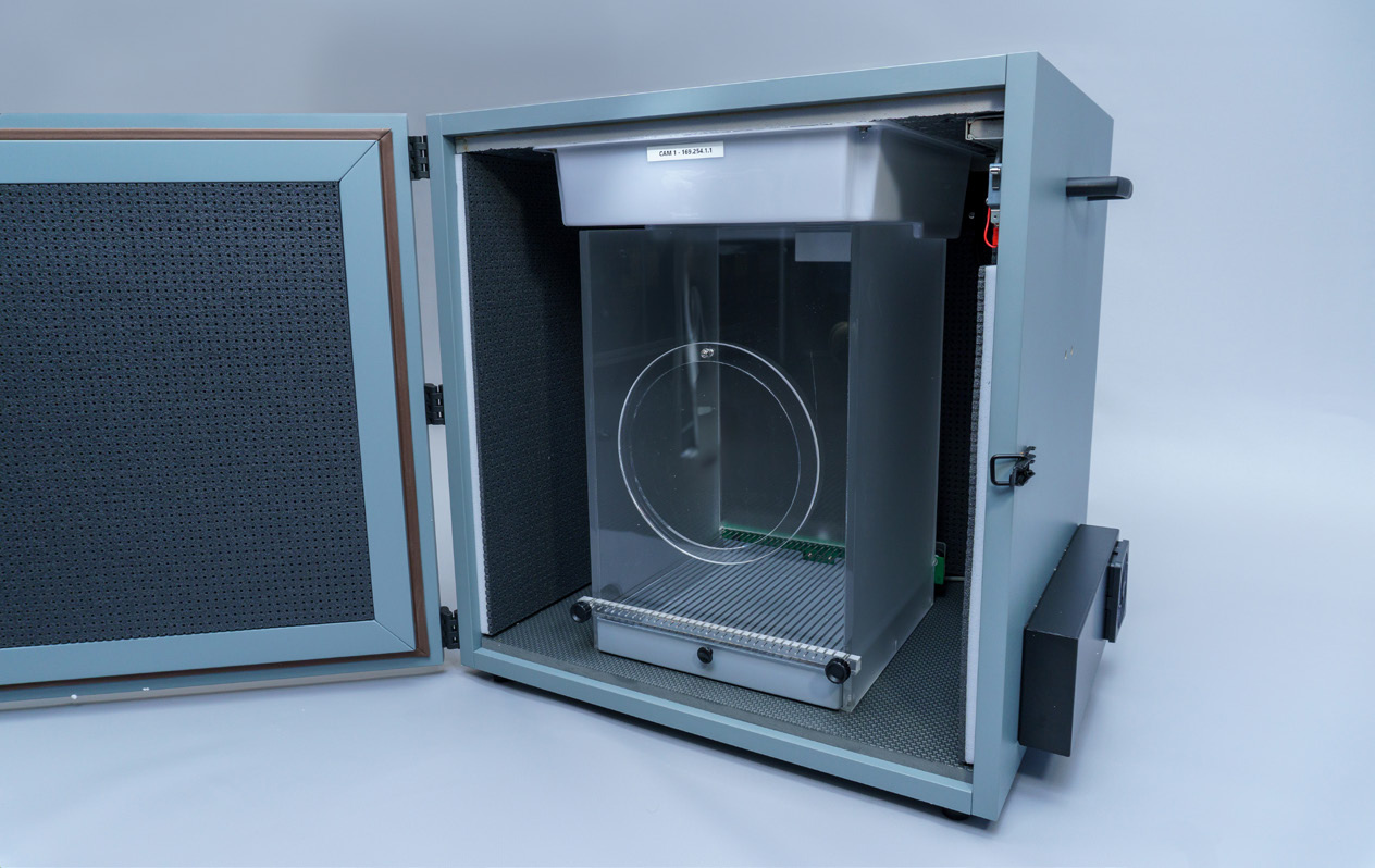 Figure 1. EthoVision XT offers an intuitive way to deliver stimuli for whichever protocol you decide to use.
Detect Freezing Behavior
Freezing behavior is one of the most important readout parameters for fear conditioning research. EthoVision XT analyzes this behavior by tracking the amount of pixel changes and the change in shape of the animal. With frame-by-frame analysis the software can easily distinguish between an active state (while the animal is grooming, moving or exploring) and an inactive state.
Pham et al. [6] used the parameter Mobility to score freezing automatically. According to the authors, EthoVision XT provides parameters that can accurately track mice and be used for automated scoring of immobility that is nearly identical to scoring by human observation.
With EthoVision XT you save a tremendous amount of time and therefore money conducting your experiments. Furthermore, with automated behavioral analysis your experiment is not biased towards the individual observer.
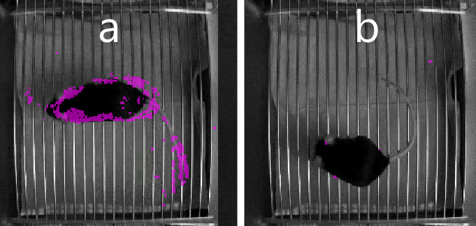 Figure 2. The Activity parameter in EthoVision XT: Purple pixels indicate the subject is active (a) or immobile (b).
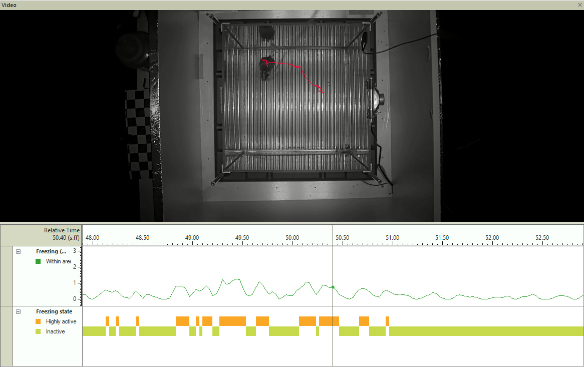 Figure 3. Activity state over time. Periods of inactive behavior could be classified as freezing.
Fear Conditioning in Practice
Effects of Gene Overexpression on Fear Learning
Glutaminase C is an enzyme involved in the process that converts glutamine to glutamate, the most important neurotransmitter in excitatory synapses. A number of neurological diseases have been associated with changes in expression of the Glutaminase C gene. Wang et al. [8] generated transgenic mice that overexpressed Glutaminase C in the brain and tested their ability to learn fear. Compared with control mice, transgenic Nes-GAC mice had a significantly lower percentage of freezing on the last day of cued fear conditioning, providing evidence that overexpression of Glutaminase C in the brain has deleterious effects on learning.
Blocking Interleukin-1 Signaling Prevents Stress-Enhanced Fear Learning
There is a general consensus that post-traumatic stress disorder involves substantial immune system dysregulation. Exposure to severe stress is associated with an increase of the signaling molecule Interleukin-1 in the hippocampus. Meghan E. Jones and collaborators (Jones et al. [9]) asked whether simply blocking the interleukin signaling was sufficient to prevent fear learning. The authors used an antagonist to block Interleukin-1 in the hippocampus. Animals that received severe stress in a specific context followed by the interleukin antagonist infusion showed significantly less freezing than animals that received the same stress followed by vehicle. Freezing was measured using the activity parameter in EthoVision XT. The authors concluded that infusion of interleukin antagonist prevented the expression of stress-enhanced fear learning.
Fear Conditioning Elicits Rapid Microstructural Changes in the Brain
There is substantial knowledge about the anatomical, functional and molecular pathways involved in fear conditioning. Establishment of persistent long-term fear memory requires structural changes in the synapses in specific regions of the brain, particularly the amygdala and hippocampal projections. Abby Ding and collaborators (Ding et al. [10]) used in vivo magnetic resonance diffusion tensor imaging (DTI) before and after fear conditioning. To record freezing behavior in the subjects, the authors used the EthoVision XT parameter Mobility which analyzes subtle changes in the contour of the animal viewed from the camera in the fear conditioning chamber. The DTI revealed rapid microstructural changes in the amygdala and hippocampus in vivo, as early as one hour after conditioning. The study confirmed that the two areas of the brain are the most important in the regulation of fear.
Depletion of Rheb GTPase Signaling Molecule Elicits Memory Defects in Mice
Ras genes encode for signaling molecules that usually work as two-state switches. If activated, a Ras molecule triggers other molecules, and ultimately determines signal pathways within the cell. Rheb (Ras homolog enriched in brain) GTPase is one of such molecules; it interacts with cell targets that are known to be implicated in life-span extension, as well as in neurodegenerative diseases such as Alzheimer's disease. However, it is not clear how Rheb GTPase participates in aging and neurodegenerative disorders. Shahani and collaborators (Shahani et al. [11]) crossed mice with Rheb with transgenic mice expressing an inducible promoter which depletes Rheb molecules. The mutant mice were then subject to contextual fear conditioning and the occurrence of freezing was recorded. Mice showed the same amount of freezing as controls. However, they performed less in spatial memory paradigms compared to controls, suggesting that Rheb depletion elicited spatial memory deficits.
References
- Wehner, J.M., Radcliffe, R.A. (2004). Cued and Contextual Fear Conditioning in Mice. Current Protocols in Neuroscience, 8.5C.1-8.5C.14. John Wiley & Sons, Inc.
- LeDoux, J.E. (2000). Emotion circuits in the brain. Annu. Rev. Neurosci, 23, 155-184.
- Blanchard, R.J., Blanchard, D.C. (1969). Crouching as an index of fear. Journal of Comparative and Physiological Psychology, 67, 370-375.
- Marren, S. (2008). Pavlovian fear conditioning as a behavioral assay for hippocampus and amygdala function: cautions and caveats. European Journal of Neuroscience, 28, 1661-1666.
- Fanselow, M.S., Sterlace, S.R. (2014). Pavlovian fear conditioning: function, cause, treatment. In: McSweeney, F.K., Murphy, E.S. (Eds.), The Wiley Blackwell Handbook of Operant and Classical Conditioning. John Wiley & Sons, Ltd., Oxford, 117-141.
- Pham, J., Cabrera, S.M., Sanchis-Segura, C., Wood, M.A. (2009). Automated scoring of fear related behavior using EthoVision software. Journal of Neuroscience Methods, 178(2), 323-326. doi:10.1016/j.jneumeth.2008.12.021.
- Anagnostaras, S.G., Wood, S.C., Shuman, T., Cai, D.J., LeDuc, A.D., Zurn, K.R., Zurn, J.B., Sage, J.R., Herrera, G.M. (2010). Automated assessment of Pavlovian conditioned freezing and shock reactivity in mice using the VideoFreeze system. Frontiers in Behavioral Neuroscience, 4, 158. doi:10.3389/fnbeh.2010.00158.
- Wang, Y., Li, Y., Zhao, R., Wu, B., Lanoha, B., Tong, Z., Peer, J., Liu, J., Xiong, H., Huang, Y., Zheng, J. (2017). Glutaminase C overexpression in the brain induces learning deficits, synaptic dysfunctions, and neuroinflammation in mice. Brain, Behavior, and Immunity, 66, 135-145.
- Jones, M.E., Lebonville, C.L., Paniccia, J.E., Balentine, M.E., Reissner, K.J., Lysle, D.T. (2018). Hippocampal interleukin-1 mediates stress-enhanced fear learning: A potential role for astrocyte-derived interleukin-1. Brain, Behavior, and Immunity, 67, 355-363.
- Ding, A.Y., Li, Q., Zhou, I.Y., Ma, S.J., Tong, G., McAlonan, G.M., Wu, E.X. (2013). MR diffusion tensor imaging detects rapid microstructural changes in amygdala and hippocampus following fear conditioning in mice. PLoS ONE, 8(1), e51704.
- Shahani, N., Huang, W.-C., Varnum, M., Page, D.T., Subramaniam, S. (2017). Forebrain depletion of Rheb GTPase elicits spatial memory deficits in mice. Neurobiology of Aging, 50, 134-143.
Download the original PDF: Fear Conditioning: How EthoVision XT Supports Your Behavioral Research (PDF)