Gait parameters as behavioral endpoints – parameters from a footprint
So here it is – the first blog in a series of three, about rodent gait analysis and what a single footprint can tell us.
Modern systems – better than ink
So what can one footprint tell you? Well, it could tell you a lot. Simply putting the paw in ink and studying the print left behind is one way to go about it, but there are far more sophisticated ways of footprint analysis. While an ink-print can give you an idea of the print area of a foot, you cannot tell how the animal is distributing his weight across its feet. It also cannot tell you the maximum surface area of a foot touching the ground during the duration of the entire footfall. Modern systems can.
Print intensity
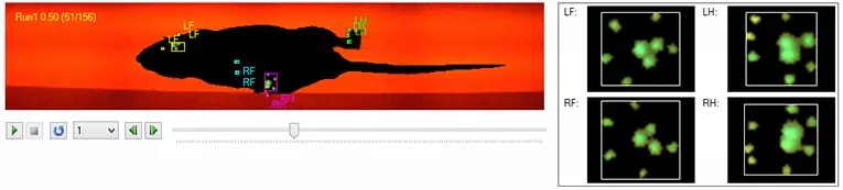
Modern systems that use light to detect a footfall can indicate the intensity of a print, which in turn can correlate with how the animal is bearing its weight. In models of conditions that affect a single limb, the animal often shows less use of the affected paw. This is reflected in a relatively lower intensity of that footprint, which is found in models of arthritis [8,9] and sciatic nerve injury [1,2,4,10].
Contact and print area
Maximum contact area and total print area are two different things that cannot be distinguished using a regular ink print. The total print area is the accumulated picture of the entire time the paw came in contact with the ground. It is what would be left behind if you had dipped that foot in ink. But cleaner. The maximum contact area is the area of the foot touching the plate during the time of maximum contact, which is usually smaller than the total print area.
Similar to the changes found in print intensity, a smaller contact area also reflects altered use of the affected limb. Again, in literature this is described in models of arthritis [8,9] and sciatic nerve injury [1,2,4,10]. Other disorders that show one or more affected paws, also show a clear reduction of print and/or contact area, such as models of ischemic stroke [5,7].
On the other hand, research on spinal cord injury reports an increased print and contact area for all four paws in a contusion injury model of spinal cord injury, and a less dramatic but still significant effect in the hind paw print areas in a dorsal cord transection model of spinal cord injury [6].
Shape of the paw
The shape of the paw, the length, and how the toes are spread, are important in research on nerve injury, arthritis and pain. These can cause deformation of the feet, altering the parts of the sole that make contact with the ground [1,2,3,4].
The sciatic nerve is a long nerve that originates in the lower back and runs down through the limbs. It branches of in the tibial and the peroneal (also called fibular) nerve. Injury to these nerves in rodents are often used as a model to study nerve regeneration, using print length, toe spread, paw angle and other gait parameters as a behavioral endpoint to measure functional recovery.
Nerve injury can cause "clawing" of the feet. In sciatic [3] or tibial nerve injury, this is often reflected in a longer print but a decreased toe spread, while damage to the peroneal nerve often results in a shorter print with a diminished toe spread. These characteristics are represented in nerve specific indexes; the sciatic functional index (SFI), the tibial functional index (TFI), and the peroneal functional index (PFI). Based on print length (PL), toe spread (TS) and intermediate toe spread (ITS), these are automatically calculated by gait analysis systems.
We can again find the advantage of using modern technology over ink dipping; the more accurate the print is (in other words: no ink smudges), the more accurate these indexes are.
Paw angle
Paw angle is the angle between the paw axis and the body axis or between the paw axis and the direction of movement (the vector). This angle can be affected by abnormalities of the muscles responsible for the placement of the paw, the nerves that innervate these muscles, or even deformation of the bones and joints of the leg belonging to the paw. Changes in paw angle are seen in models of arthritis, ataxia, stroke, and spinal cord injury.
Automated systems
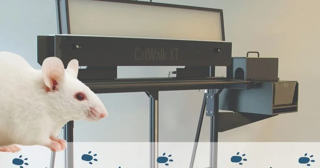
All the parameters and indexes mentioned in this blog can automatically be detected, measured, and calculated with gait analysis systems such as CatWalk XT. Now imagine adding inter-paw parameters such as the distance between paws, or the length of a stride. And what if you added time to your analysis? Stay tuned for the other blogs in this series…
RESOURCES
Read more about CatWalk XT
Find out how CatWalk XT is used in a wide range of studies and how it can elevate your research!
- Free white papers and case studies
- Customer success stories
- Recent blog posts
Resources
arrow_forward
Sign up for more blogs!
If you don't want to miss out on the next two blogs in this series, sign up here to be notified when they are online. (If you have already signed up previously, you don't need to do so again.)
Other blogs in this series
Other blogs in this series are now online:
References
- Bozkurt, A.; Deumens, R.; Scheffel, J.; O'Dey, D.M.; Weis, J.; Joosten, E.A.; Führman, T.; Brook, G.A.; Pallua, N. (2008). CatWalk gait analysis in assessment of functional recovery after sciatic nerve injury. Journal of Neuroscience Methods, 173, 91-98.
- Bozkurt, A.; Scheffel, J.; Brook, G.A.; Joosten, E.A.; Suschek, C.V.; O'Dey, D.M.; Pallua, N.; Deumens, R. (2011). Aspects of static and dynamic motor function in peripheral nerve regeneration: SSI and CatWalk gait analysis. Behavioural Brain Research, 219, 55-62.
- Costa, L.M.; Simões, M.J.; Maurício, A.C.; Varejão, A.S.P. (2009). Methods and protocols in peripheral nerve regeneration experimental research: Part IV – kinematic gait analysis to quantify peripheral nerve regeneration in the rat. International Review of Neurobiology, 87, 127-139.
- Deumens, R.; Jaken, R.J.P.; Marcus, M.A.E.; Joosten, E.A.J. (2007). The CatWalk gait analysis in assessment of both dynamic and static gait changes after adult rat sciatic nerve resection. Journal of Neuroscience Methods, 164, 120-130.
- Encarnacion, A.; Horie, N.; Keren-Gill, H.; Bliss, T.M.; Steinberg, G.K.; Shamloo, M. (2011). Long-term behavioral assessment of function in an experimental model for ischemic stroke. Journal of Neuroscience Methods, 196, 247-257.
- Hamers, F.P.T.; Lankhorst, A.J.; Van Laar, T.J.; Veldhuis, W.B.; Gispen, W.H. (2001). Automated quantitative gait analysis during overground locomotion in the rat: its application to spinal cord contusion and transection injuries. Journal of Neurotrauma, 18, 187-201.
- Hetze, S.; Römer, C.; Teufelhart, C.; Meisel, A.; Engel, O. (2012). Gait analysis as a method for assessing neurological outcome in a mouse model of stroke. Journal of Neuroscience Methods, 206, 7-14.
- Möller, K.Ä.; Berge, O.-G.; Hamers, F.P.T. (2008). Using the CatWalk method to assess weight-baring and pain behaviour in walking rats with ankle joint monoarthritis induces by carrageenan: effects of morphine and rofecoxib. Journal of Neuroscience Methods, 174, 1-9.
- Pinho, D.; Morato, M.; Couto, M.R.; Marques-Lopes, J.; Tavares, I.; Albino-Teixeira. A. (2011). Does chronic pain alter the normal interaction between cardiovascular and pain regulatory systems? Pain modulation in the hypertensive-monoarthritic rat. The Journal of Pain, 12, 194-204.
- Sheu, M.L.; Cheng, F.-C.; Su, H.-L.; Chen, Y.-J.; Chen, C.-J.; Chiang, C.-M.; Chiu, W.-T.; Sheehan, J.; Pan, H.-C. (2012). Recruitment by SDF-1α of CD34-positive cells involved in sciatic nerve regeneration. Journal of Neurosurgery, 116, 432-444.