Rodents such as rats and mice are powerful animal models for studying the mechanisms of behavior. Recent advances in imaging technology have enabled scientists to study the underlying neuronal networks responsible for various behaviors in rodents. One of the most powerful methods is calcium imaging, which allows researchers to observe activity patterns within neuronal networks and track changes in neuronal responses associated with different behaviors. In this blog, we will explore what this technique is, and how it can advance your behavioral research.
What is Calcium Imaging?
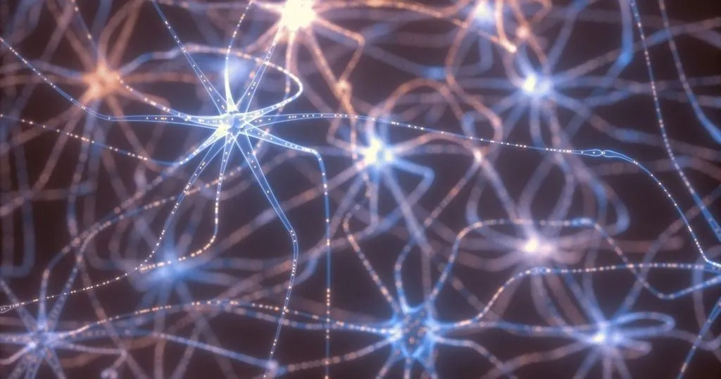
Calcium imaging enables researchers to observe and measure the activity of individual neurons in real-time, offering valuable insights into complex behaviors and the functioning of deep brain regions like the prefrontal cortex or hippocampus. Calcium ions (Ca2+) are essential signaling molecules involved in numerous cellular processes, including neuronal communication, muscle contraction, and gene expression. Changes in calcium ion concentrations indicate cellular activity, allowing researchers to decode neural activity associated with specific behaviors by monitoring these shifts within neurons.
The basic principles of calcium imaging
Calcium imaging involves using specialized fluorescent dyes or genetically encoded calcium indicators to detect changes in calcium ion concentrations. There are several types of calcium indicators that emit fluorescence when they bind to calcium ions. These indicators can be synthetic dyes or genetically encoded proteins. The most commonly used synthetic dyes include Fluo-4, Rhod-2, and Oregon Green BAPTA, while genetically encoded indicators include GCaMP and R-GECO.
- The chosen calcium indicator is introduced into the target cells. For example, in the case of neurons, the indicator can be injected into the neurons. They are loaded via diffusion through a patch pipette, or expressed using viral vectors for genetically encoded indicators.
- Once the indicator is inside the cells, it is excited by a specific wavelength of light, typically using a fluorescent microscope. When the indicator molecule binds to calcium ions, its fluorescence properties change. This change can be detected using a sensitive camera or photodetector.
- The microscope captures images of the cells over time, recording the changes in fluorescence intensity. The intensity of fluorescence emitted by the indicator is proportional to the concentration of calcium ions in the cell. An increase in calcium ion concentration leads to an increase in fluorescence intensity, while a decrease leads to a decrease in intensity.
- The collected fluorescence intensity data can be analyzed to extract information about cellular activity. Researchers can quantify the frequency, amplitude, and duration of calcium transients, which correspond to specific cellular events. For instance, in neurons, calcium transients often correspond to action potentials or other types of electrical activity.
- Calcium imaging allows researchers to visualize both spatial and temporal patterns of calcium activity. By observing multiple cells simultaneously, researchers can gain insights into how cells interact within a network.
However, calcium imaging has some limitations, including potential phototoxicity (damage to cells due to intense light exposure), photobleaching (loss of fluorescence signal over time), and potential interference of the indicator with cellular processes. Additionally, the indicator's affinity for calcium ions and its rate of binding/unbinding can impact the accuracy of the measurements.
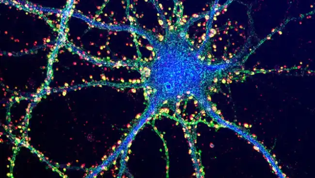
Image credit: Society for Neuroscience
Calcium imaging and studying (rodent) behavior
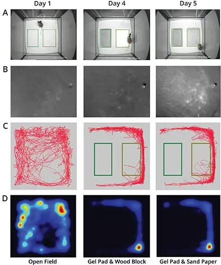
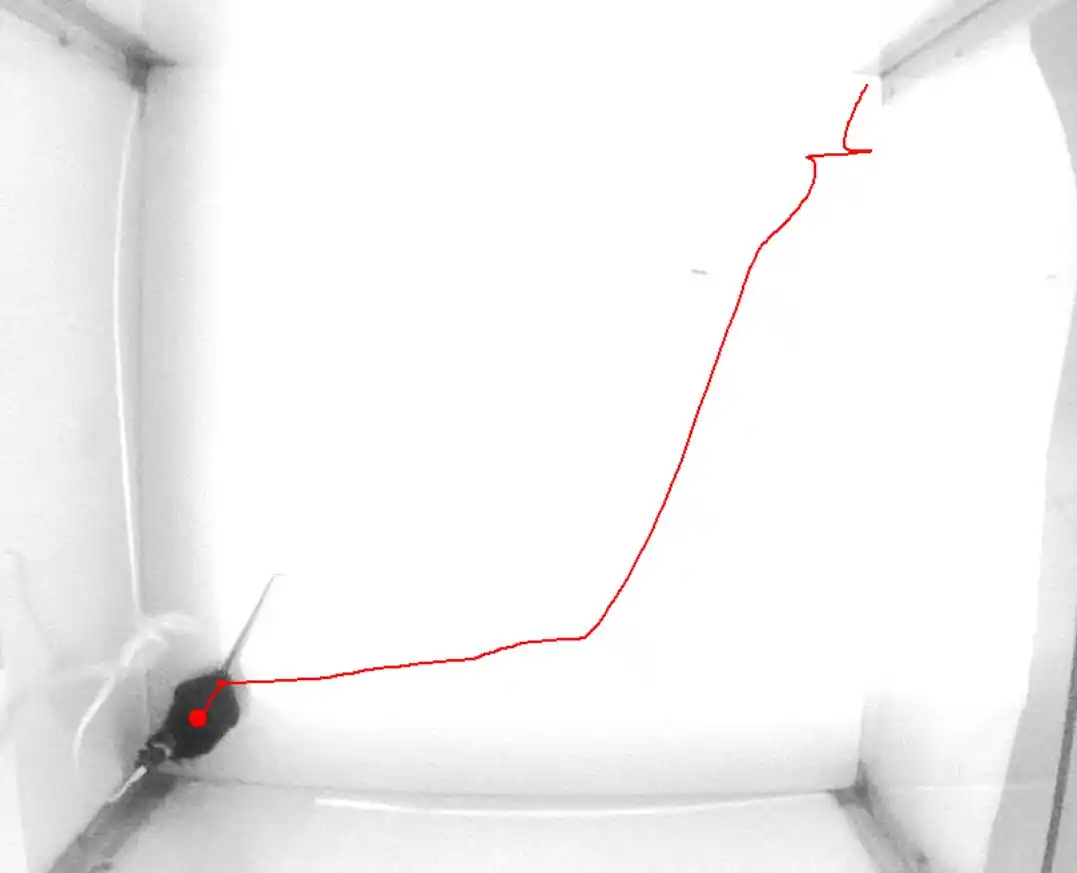
To analyze and interpret these large amounts of data from within the brain, and link them to behavior, researchers often rely on sophisticated behavioral software tools, such as Noldus' EthoVision XT. This software platform enables researchers to precisely track and analyze the behavior of rodents during calcium imaging experiments.
Examples of calcium imaging behavioral studies
- One such study, published in the journal Nature Communications utilized calcium imaging techniques to investigate the role of serotonergic neurons in social behaviors in mice. The researchers employed a combination of calcium imaging and behavioral analysis to study how activity in specific serotonergic neuron populations influenced social interaction in mice. This study revealed the importance of serotonergic neurons in modulating social behaviors and provided valuable insights into the neural mechanisms underlying social interaction.
- Another example is a study published in the journal Cell Reports that examined the neural activity in the amygdala of mice when faced with a threat. The researchers describe the presence of neurons expressing calcitonin gene-related peptide (CGRP) in two specific brain regions: the parvocellular subparafascicular nucleus in the thalamus and the external lateral parabrachial nucleus in the brainstem. These neurons are found to respond to threat cues from multiple sensory modalities and convey negative emotional valence signals to different parts of the amygdala. The study shows that both populations of CGRP-expressing neurons and their connections to the amygdala are crucial for the perception of multi-sensory threats and the formation of aversive memories. This finding suggests a unified neural pathway for innate threat perception, which could have implications for the development of therapeutic approaches for disorders related to innate fear responses.
VIDEO TRACKING SOFTWARE
Track behavior with EthoVision XT
Analyze rodent behavior during calcium imaging experiments with precision and ease.
Learn more
arrow_forward
Benefits of calcium imaging and studying behavior
- One of the key benefits of calcium imaging is its ability to capture the dynamics of neuronal activity in real-time. This enables researchers to observe how specific behaviors activate distinct populations of neurons and track their temporal dynamics. By analyzing the neural activity associated with different behavioral states, such as exploration, social interaction, or fear response, researchers can gain a better understanding of the neural mechanisms underlying these complex behaviors.
- In addition, calcium imaging allows for the investigation of deep brain regions, which play critical roles in various behaviors and brain functions. By imaging these deep brain regions, such as the prefrontal cortex or hippocampus, researchers can reveal how neural activity in these regions contributes to specific behavioral outputs.
- Furthermore, calcium imaging has promising applications in evaluating therapeutic efficacy. Researchers can use calcium imaging to assess how neuronal activity is altered by different interventions, such as drug administration or deep brain stimulation. By longitudinally monitoring the neural activity in targeted brain regions, researchers can determine the impact of these interventions on neural circuit dynamics and behavior.
Other species used in calcium imaging studies
In addition to mice and rats, other species are commonly used in calcium imaging research to study behavior. These species provide unique opportunities to explore a wide range of behaviors in various contexts. Here are some examples:
Zebrafish
Zebrafish are a popular model organism for studying behavior due to their transparent embryos and larvae, which allows for non-invasive imaging of neural activity. Their small size and rapid development make them well-suited for high-throughput behavioral assays. Calcium imaging studies in larval zebrafish has, for example, potentially uncovered personalized epilepsy treatments.
Drosophila (fruit flies)
Fruit flies are a widely used model organism in neuroscience research. They have a well-characterized nervous system and exhibit a diverse range of behaviors, including courtship, locomotion, learning, and memory. Calcium imaging in Drosophila provides insights into the underlying neural circuitry involved in these behaviors. Check out this study by Patel et al (2022) that employs calcium imaging and behavioral analysis with EthoVision XT to understand how animals would sense innocuous and/or potentially harmful stimuli
C. elegans (roundworms)
Despite their small size and simple nervous system, C. elegans (roundworms) offer unique advantages for behavioral studies. However, calcium imaging in C. elegans is challenging due to the difficulties associated with immobilizing the organism. Check out this recent study, in which the authors have developed a new method to immobilize worms by trapping them in sodium alginate gel.
Zebra Finch
Songbirds such as the zebra finch are known for their complex vocal learning behaviors. This study focuses on understanding how zebra finches learn and produce their complex songs. Using calcium imaging and two-photon microscopy, the authors examined the process that extends from higher-order brain centers to muscle movements. They monitored ensemble activity during singing to establish that a crucial forebrain structure known as HVC contains premotor neurons that become active at specific time points during song production.
HOME CAGE OBSERVATION
Study behavior in the PhenoTyper
Integrate calcium imaging with automated home cage monitoring for comprehensive behavioral analysis.
Discover PhenoTyper
arrow_forward
Conclusion
In summary; calcium imaging, by using optical imaging technologies such as single and two-photon imaging of calcium indicators, will continue to play an important role for measuring behavior and its neural correlates in rodents. This technique's ability to measure neuronal activity with high spatial resolution stands out; allowing researchers to not only measure the activity of individual neurons, but also to pinpoint the specific neurons and brain regions involved in a particular behavior.
These findings have implications for understanding the neural basis of behavior and may have potential therapeutic applications in the future. With the advancement of technology and software tools, such as Noldus' EthoVision XT and the PhenoTyper, which can seamlessly integrate with calcium imaging systems, giving researchers the ability to analyze and interpret calcium imaging data in freely moving rats and mice, further enhancing our understanding of behavior.